The angle between the operative defect and the primary lobe is 45 degrees.
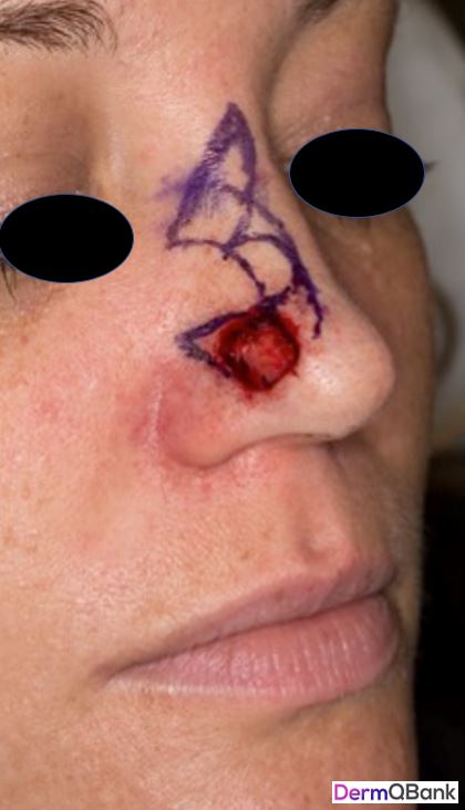
The flap design shown is that of a bilobed transposition flap of the nose, which is the flap of choice for many dermatologic surgeons for defects < 1.0 cm (in patients with more mobile, non sebaceous skin, up to 1.5 cm) in diameter on the nasal tip and supratip.
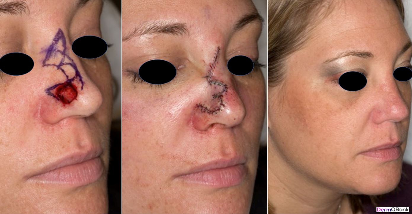
The design and execution of the bilobed flap are crucial in order to achieve a superior aesthetic repair, and without great attention to detail, this flap can lead to pincushioning and alar asymmetry.
The goal of the flap is to direct all tension to the horizontal axis such that the flap simply drapes into the postoperative defect (typically from micrographic dermatologic surgery). Where possible, the secondary lobe of the flap (which is triangulated) is designed to be oriented vertically.
The angle between each lobe of the flap should measure 45 to 50 degrees, which makes for a 90 to 100 degree total arc of transposition between the defect and the secondary lobe.
Breakdown of a properly designed and executed flap (Goldman, Glen. Facial Flap Surgery. McGraw-Hill. 2013):
- Deepen the operative defect on the nasal tip/supratip to perichondrium
- Determine the apex of the standing tissue cone (angled superolaterally for lateral-based bilobed flap to minimize alar distortion)
- Determine flap pivot point (which is not necessarily the apex of the standing cone) and delineate an arc equal in length to the distance from the pivot point to the distal-most point of operative defect. Create a 90 degree angle
- Design two lobes equal is size with 45 degree angles from within the arc already created (see above), making sure to triangulate the secondary lobe (aligned as vertically as possible), as this portion will be closed linearly
- Many remove the standing cone at this point (down to the nasalis superiorly and perichondrium inferiorly), and thereafter, carefully undermine (widely) the flap above the perichondrium and periosteum. With care, the nasalis arterial branch accompanying the nasalis muscle can be preserved. And with the flap elevated submuscularly, the nasociliary nerve may be preserved, which avoids loss of sensory to the nasal tip
- The standing rule for transposition flaps is to close the secondary (rhombic) or tertiary (bilobed) defects first, given the maximum tension at these points. But in nasal bilobed flaps, some choose to close the primary defect first
Clinical pearl: The angle between each lobe of the bilobed flap should measure about 45 degrees, which makes for a total arc angle of 90 degrees.